The patient was a 36-year-old man who experienced a rapid decline in vision approximately two months before being seen at our practice. He was found to have an acute medical condition that required hospitalization. The condition was treated, and his vision improved but did not fully recover. At his initial presentation to our practice, the visual acuity was 20/50 (pinhole, 20/40) OD and 20/200 (pinhole, 20/70) OS. Two months later, visual acuity was 20/30-1 OD and 20/60-1 OS.
What was the most likely diagnosis?
A. Purtscher's retinopathy
B. Multiple myeloma
C. Hypertensive retinopathy
D. Diabetic retinopathy
Answer
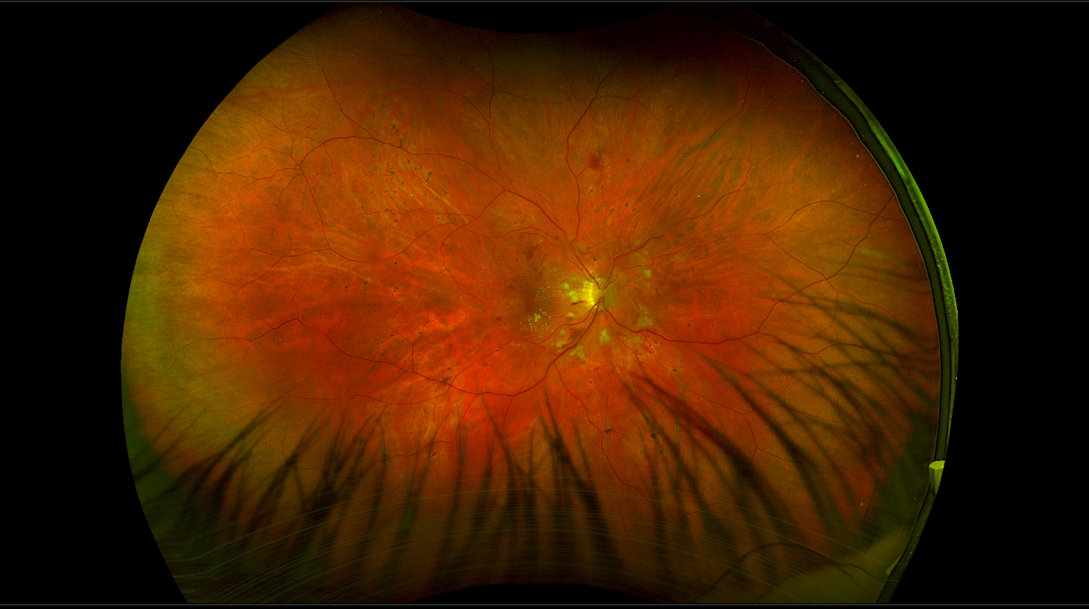
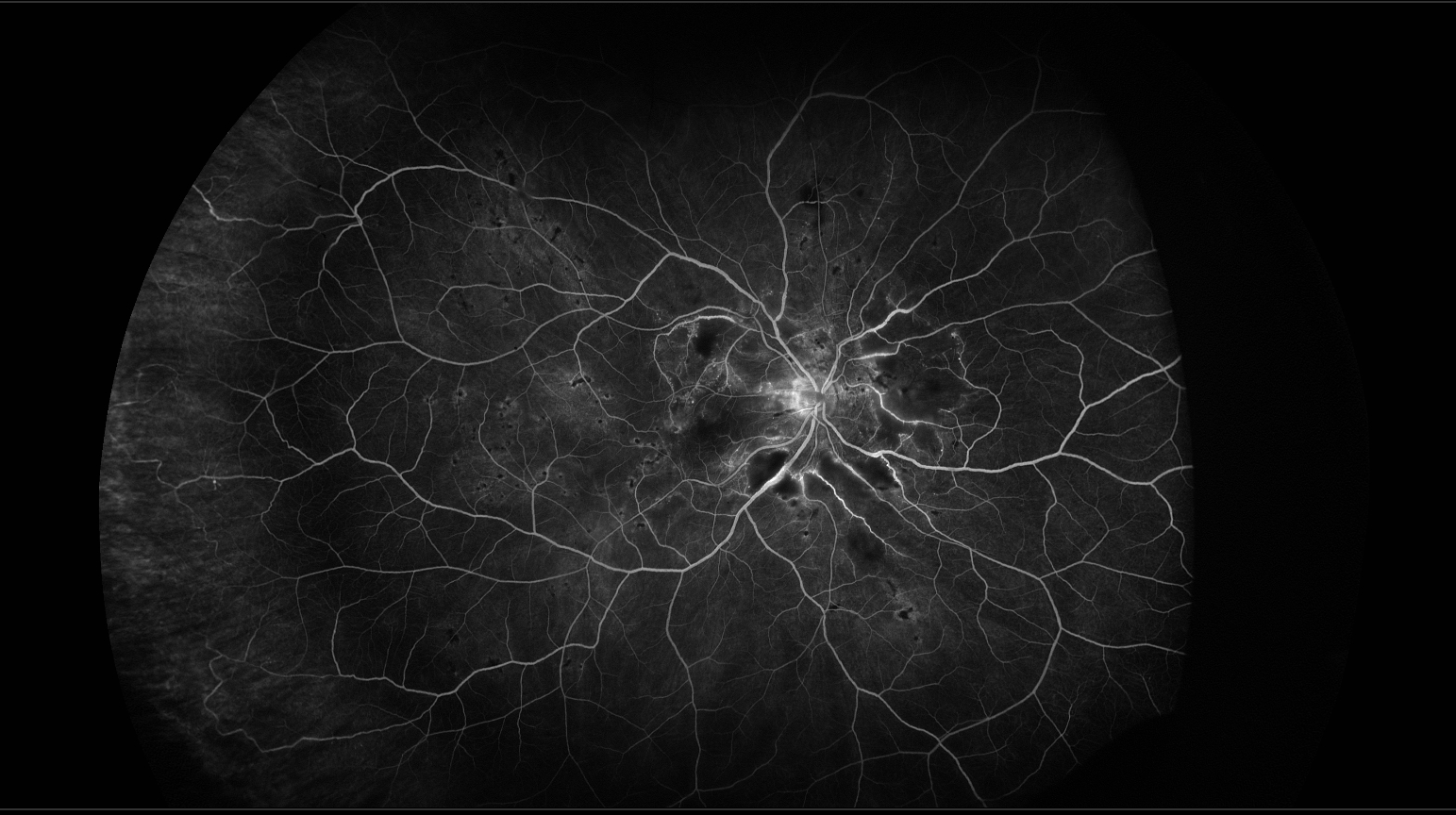
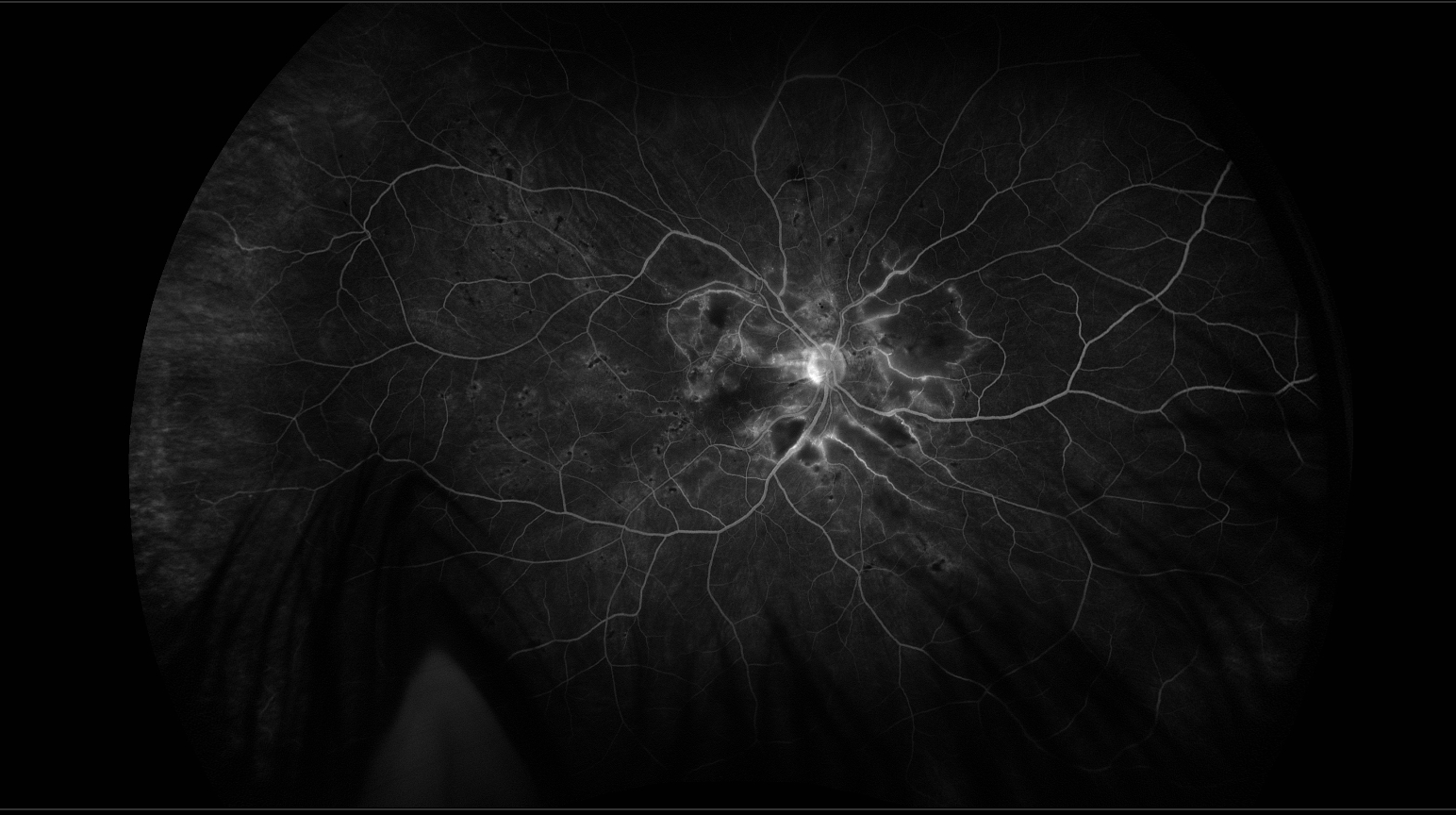
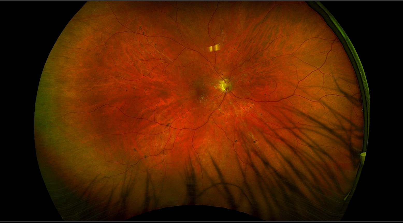
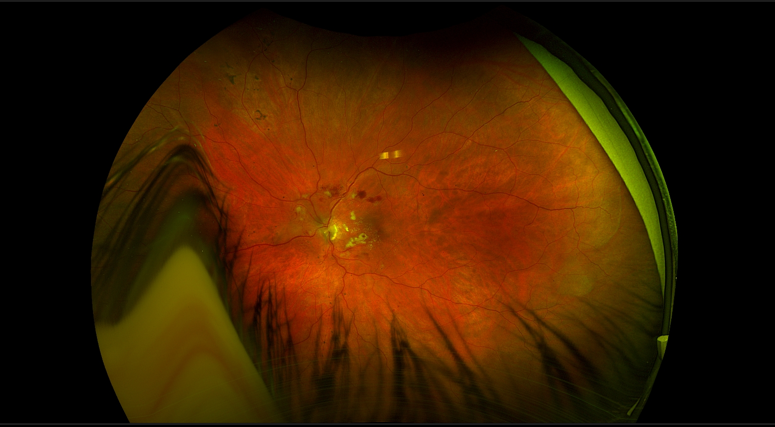
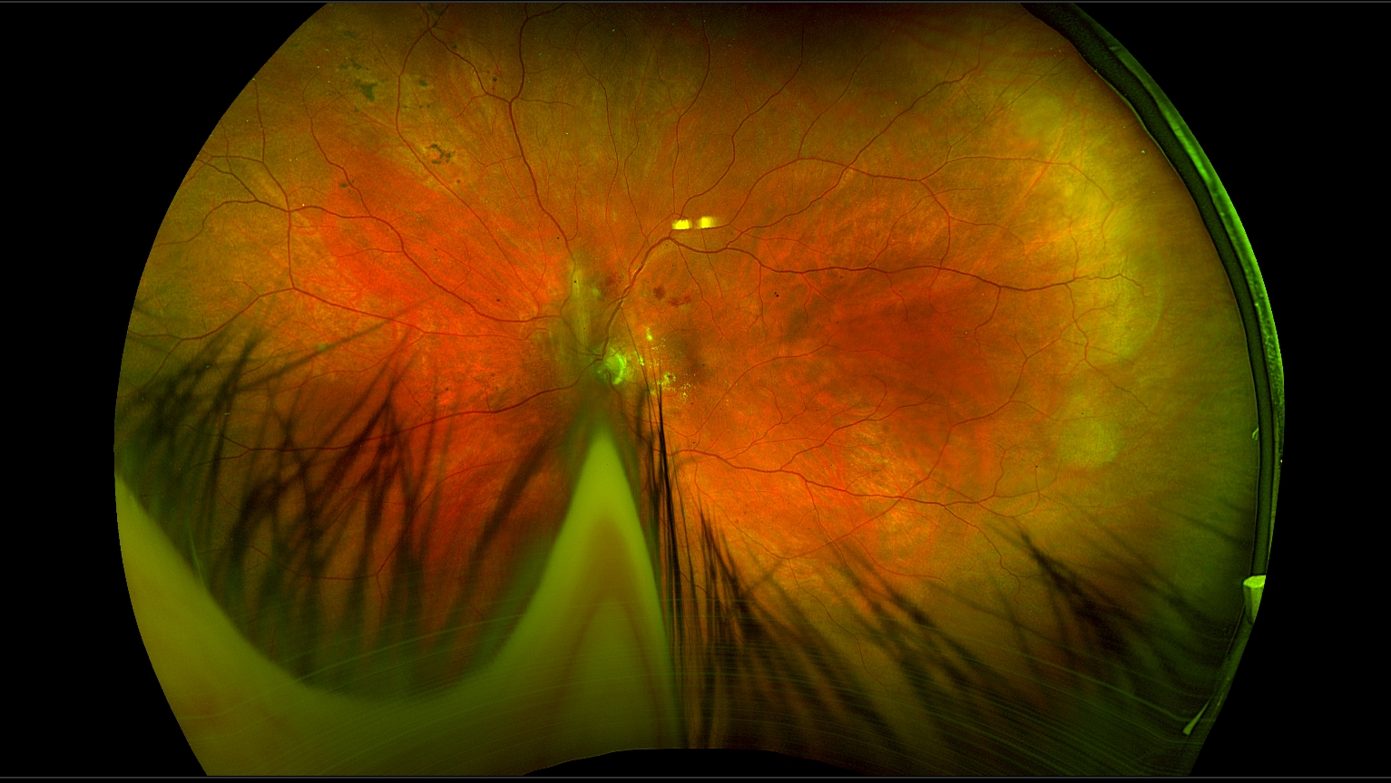
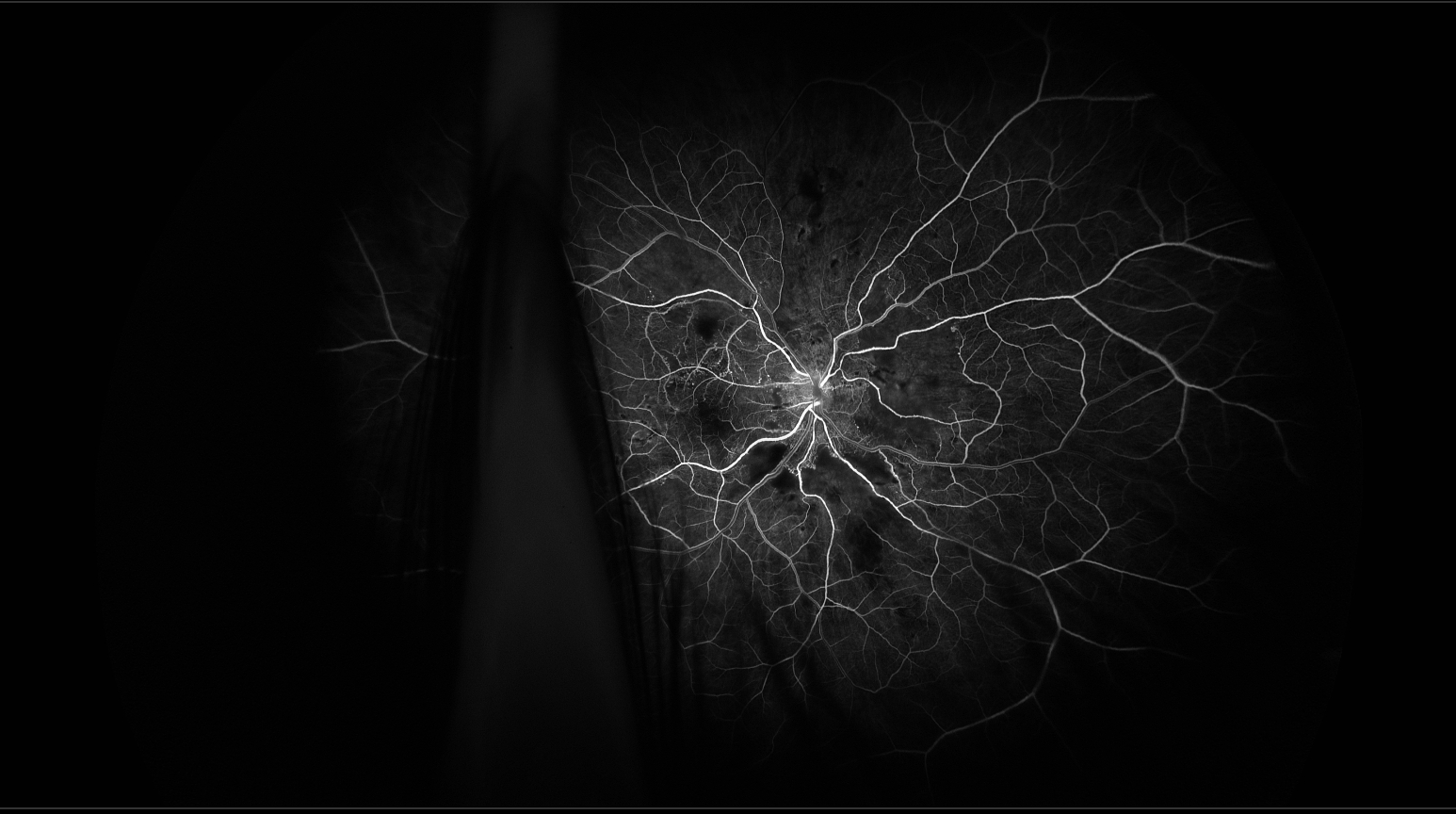
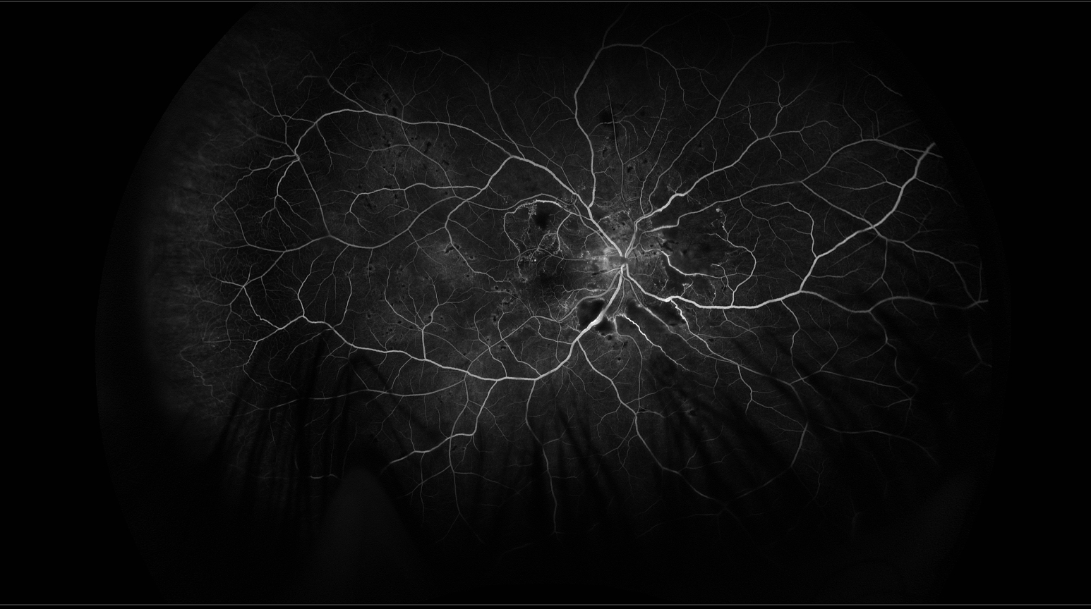
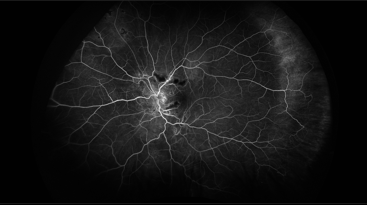
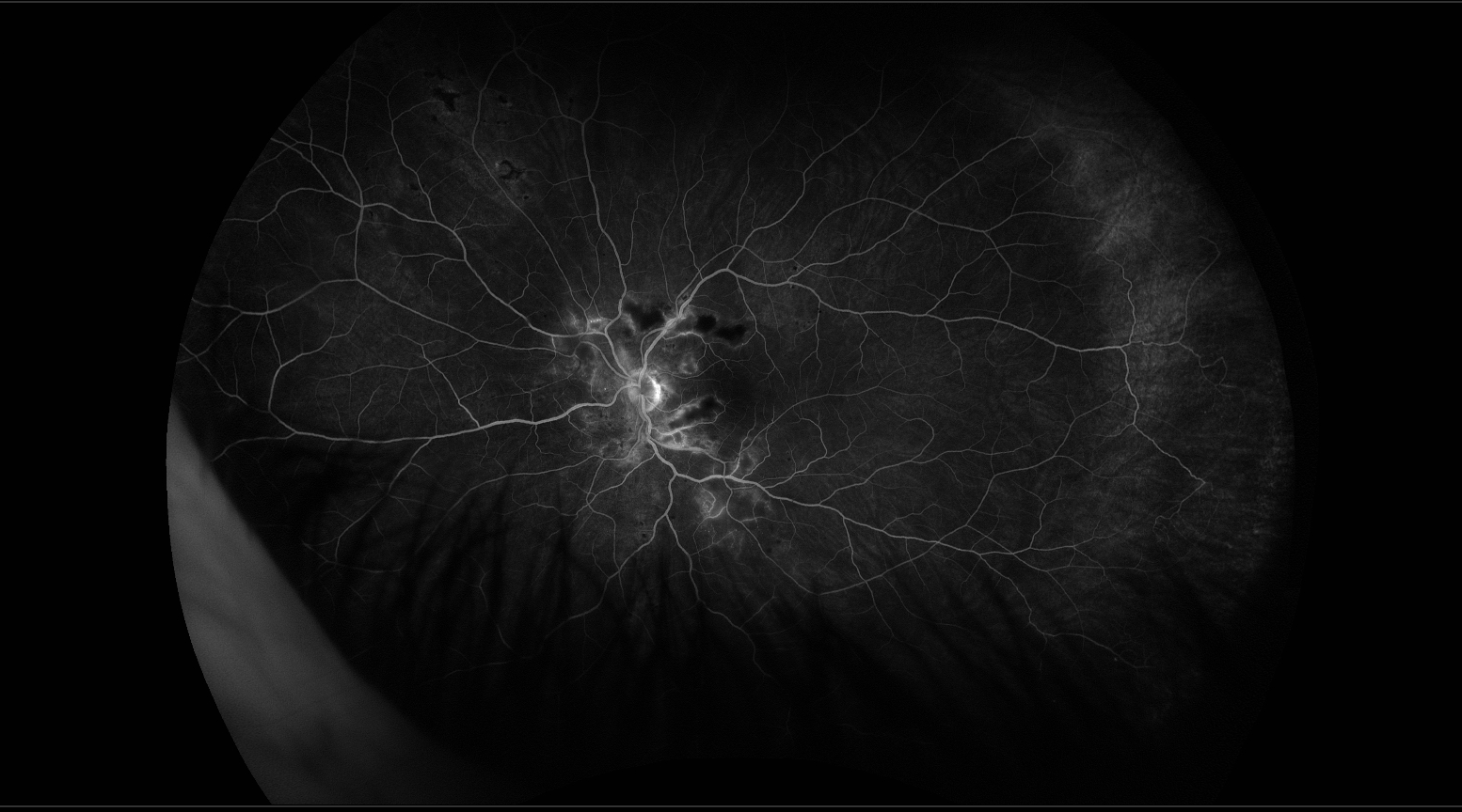
The fundus photographs show hemorrhages, cotton-wool spots, and hard exudates in both eyes. There is no disc edema in either eye. Numerous small, deep hyperpigmented spots are present outside the macula in both eyes; these appear to be Elschnig spots. There are also deep whitish streaks that appear to be Siegrist streaks. Fluorescein angiography of the right eye shows normal filling times. There is nonperfusion, most prominent in the macula and nasal to the disc. The left eye appears to have more severe macular nonperfusion than the right eye. There is late leakage from blood vessels in the areas of nonperfusion.
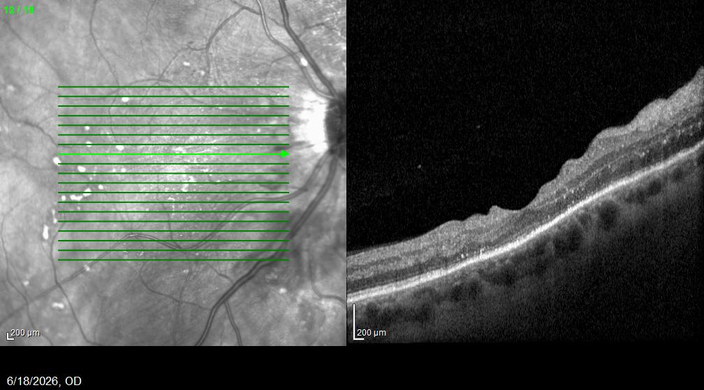
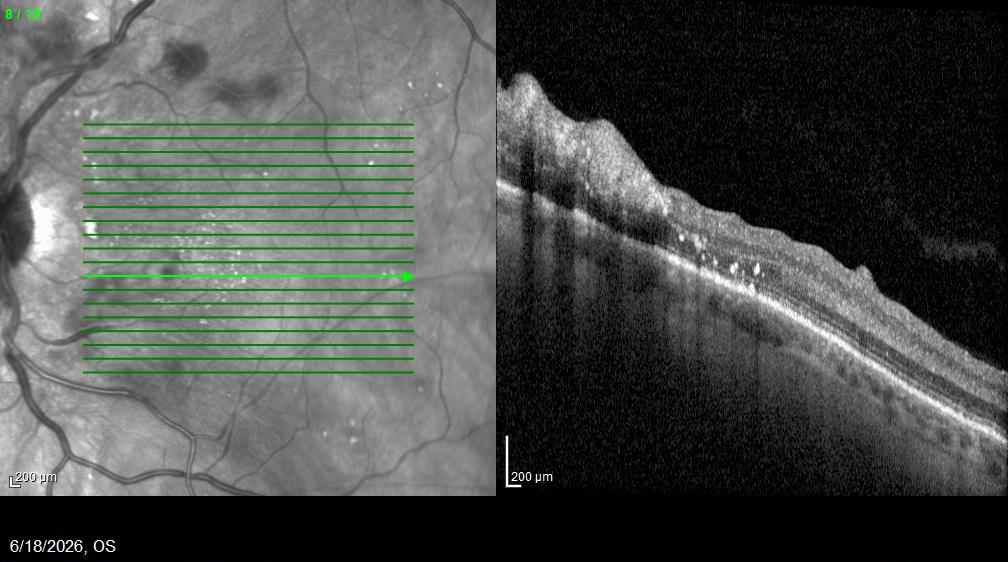
OCT of both eyes shows inner retinal edema, primarily nasal to the fovea. There are surface undulations caused by the edema and cotton-wool spots, with no epiretinal membrane in either eye. Both eyes have small hyperreflective spots in the inner and outer retina. There is also disruption of the ellipsoid zone, greater in the right eye than the left. The right eye has choroidal thickening and very large choroidal blood vessels (pachyvessels). Two months later, both eyes had substantially fewer cotton-wool spots and hemorrhages, but there was only a modest reduction in the number of hard exudates.
These findings are consistent with severe hypertensive retinopathy. The patient had been admitted to the hospital two months earlier for blood pressure control; he reported that his systolic blood pressure was 288 mm Hg at the time. In our office, his blood pressure was 150/90 mm Hg. Patients with hypertensive retinopathy can experience permanent vision loss from retinal nonperfusion, and malignant hypertension can cause optic nerve damage. Our patient did not have disc edema at presentation or subsequent disc pallor. Rarely, patients with malignant hypertension are also at risk of developing neovascularization.
Small blood vessels are damaged by the markedly elevated pressure associated with acute hypertension. Cotton-wool spots result from occlusion of the superficial retinal capillaries, whereas Elschnig spots are secondary to infarction of the choriocapillaris. Damage to the choriocapillaris produces changes in the overlying retinal pigment epithelium (RPE), accounting for the areas of deep hyperpigmentation. Elschnig spots are characteristically associated with hypertension and are more frequently found in younger patients, whose blood vessels have greater elasticity and more active vasomotor responses, allowing them to react intensely to sudden, severe elevations in blood pressure.
Siegrist streaks are caused by fibrosis of the choroidal blood vessels. In addition to severe hypertension, they can occur in inflammatory vasculopathies such as giant cell arteritis. The hyperreflective intraretinal spots found in patients with hypertensive retinopathy may represent hard exudates, proteins, and areas of microglial activation. Disruption of the ellipsoid zone and choroidal thickening with pachyvessels are also frequent findings in hypertensive retinopathy and may be related to impaired choriocapillaris and choroidal blood flow.
Several features distinguish hypertensive retinopathy from the other options. Purtscher's retinopathy typically presents with cotton-wool spots and small-vessel occlusions, with far fewer hemorrhages than were seen in our patient. Hard exudates are not typically present in this condition. The most common causes of Purtscher's retinopathy include head trauma, blunt chest trauma, long-bone fractures, and pancreatitis.
Patients with multiple myeloma can develop hyperviscosity syndrome, which can lead to numerous hemorrhages and cotton-wool spots; however, hard exudates would not be expected. There is also often venous dilation and tortuosity, which were not present in our patient.
Our patient differed from patients with diabetic retinopathy in that there were no evident microaneurysms, and the sudden bilateral onset was atypical. Patients with more severe diabetic retinopathy often have significant peripheral nonperfusion, whereas our patient's nonperfusion was largely posterior to the equator.
The fundus photographs show hemorrhages, cotton-wool spots, and hard exudates in both eyes. There is no disc edema in either eye. Numerous small, deep hyperpigmented spots are present outside the macula in both eyes; these appear to be Elschnig spots. There are also deep whitish streaks that appear to be Siegrist streaks. Fluorescein angiography of the right eye shows normal filling times. There is nonperfusion, most prominent in the macula and nasal to the disc. The left eye appears to have more severe macular nonperfusion than the right eye. There is late leakage from blood vessels in the areas of nonperfusion.
OCT of both eyes shows inner retinal edema, primarily nasal to the fovea. There are surface undulations caused by the edema and cotton-wool spots, with no epiretinal membrane in either eye. Both eyes have small hyperreflective spots in the inner and outer retina. There is also disruption of the ellipsoid zone, greater in the right eye than the left. The right eye has choroidal thickening and very large choroidal blood vessels (pachyvessels). Two months later, both eyes had substantially fewer cotton-wool spots and hemorrhages, but there was only a modest reduction in the number of hard exudates.
These findings are consistent with severe hypertensive retinopathy. The patient had been admitted to the hospital two months earlier for blood pressure control; he reported that his systolic blood pressure was 288 mm Hg at the time. In our office, his blood pressure was 150/90 mm Hg. Patients with hypertensive retinopathy can experience permanent vision loss from retinal nonperfusion, and malignant hypertension can cause optic nerve damage. Our patient did not have disc edema at presentation or subsequent disc pallor. Rarely, patients with malignant hypertension are also at risk of developing neovascularization.
Small blood vessels are damaged by the markedly elevated pressure associated with acute hypertension. Cotton-wool spots result from occlusion of the superficial retinal capillaries, whereas Elschnig spots are secondary to infarction of the choriocapillaris. Damage to the choriocapillaris produces changes in the overlying retinal pigment epithelium (RPE), accounting for the areas of deep hyperpigmentation. Elschnig spots are characteristically associated with hypertension and are more frequently found in younger patients, whose blood vessels have greater elasticity and more active vasomotor responses, allowing them to react intensely to sudden, severe elevations in blood pressure.
Siegrist streaks are caused by fibrosis of the choroidal blood vessels. In addition to severe hypertension, they can occur in inflammatory vasculopathies such as giant cell arteritis. The hyperreflective intraretinal spots found in patients with hypertensive retinopathy may represent hard exudates, proteins, and areas of microglial activation. Disruption of the ellipsoid zone and choroidal thickening with pachyvessels are also frequent findings in hypertensive retinopathy and may be related to impaired choriocapillaris and choroidal blood flow.
Several features distinguish hypertensive retinopathy from the other options. Purtscher's retinopathy typically presents with cotton-wool spots and small-vessel occlusions, with far fewer hemorrhages than were seen in our patient. Hard exudates are not typically present in this condition. The most common causes of Purtscher's retinopathy include head trauma, blunt chest trauma, long-bone fractures, and pancreatitis.
Patients with multiple myeloma can develop hyperviscosity syndrome, which can lead to numerous hemorrhages and cotton-wool spots; however, hard exudates would not be expected. There is also often venous dilation and tortuosity, which were not present in our patient.
Our patient differed from patients with diabetic retinopathy in that there were no evident microaneurysms, and the sudden bilateral onset was atypical. Patients with more severe diabetic retinopathy often have significant peripheral nonperfusion, whereas our patient's nonperfusion was largely posterior to the equator.