Answer
Choroidal macrovessel
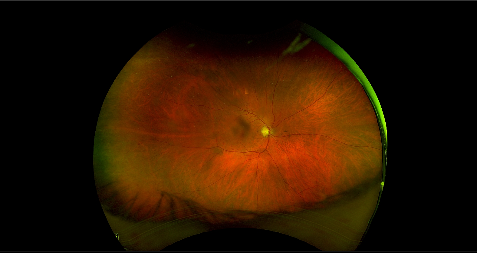
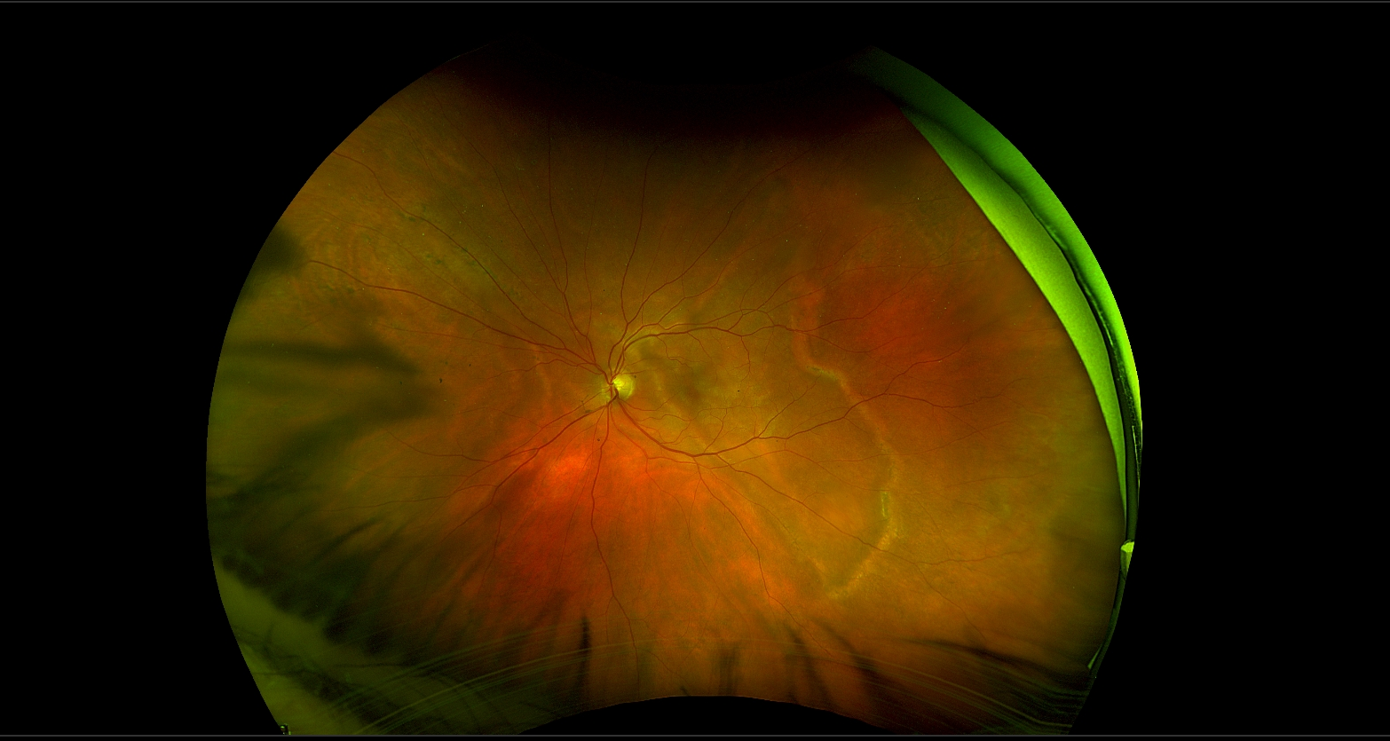
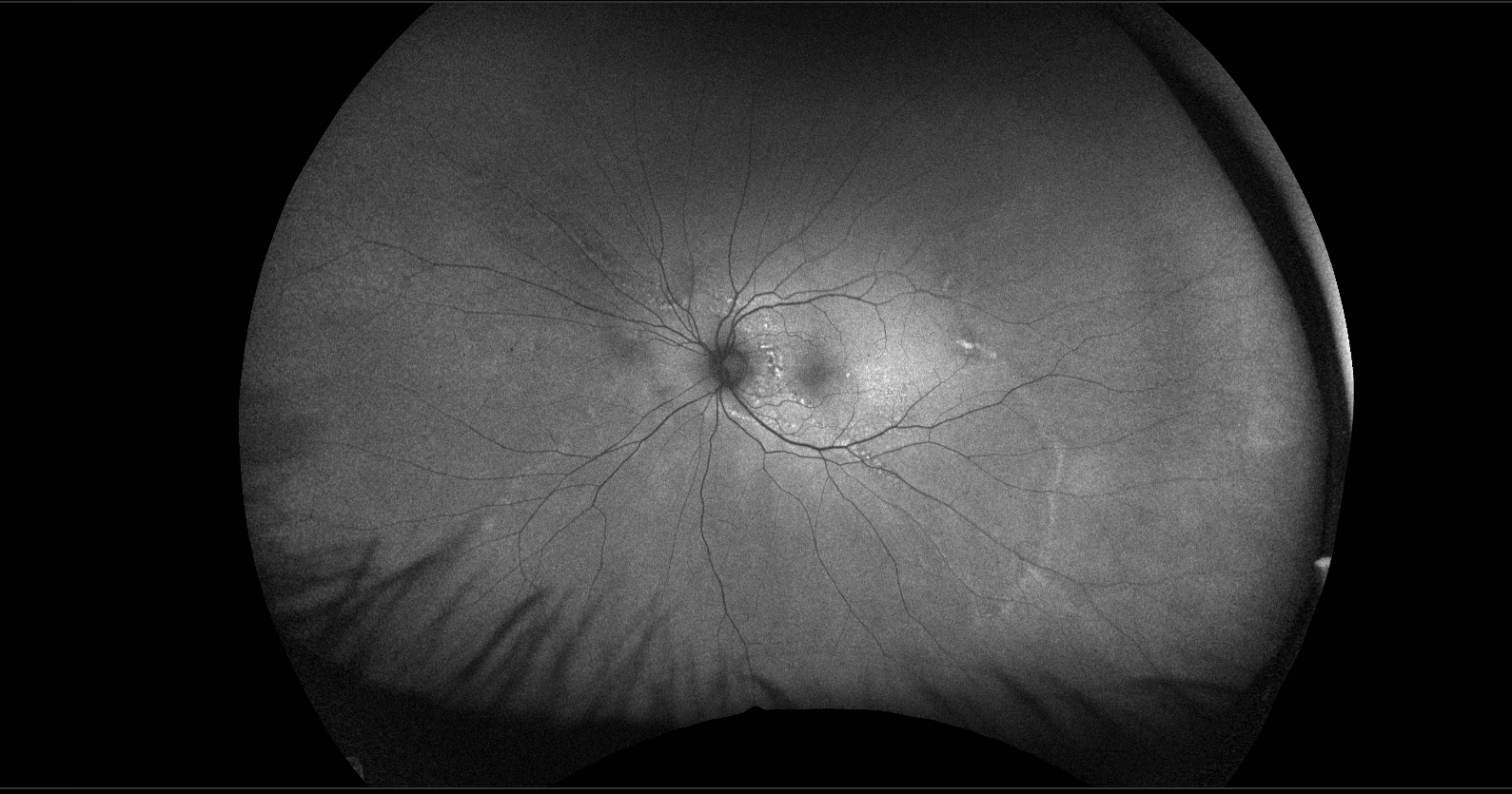
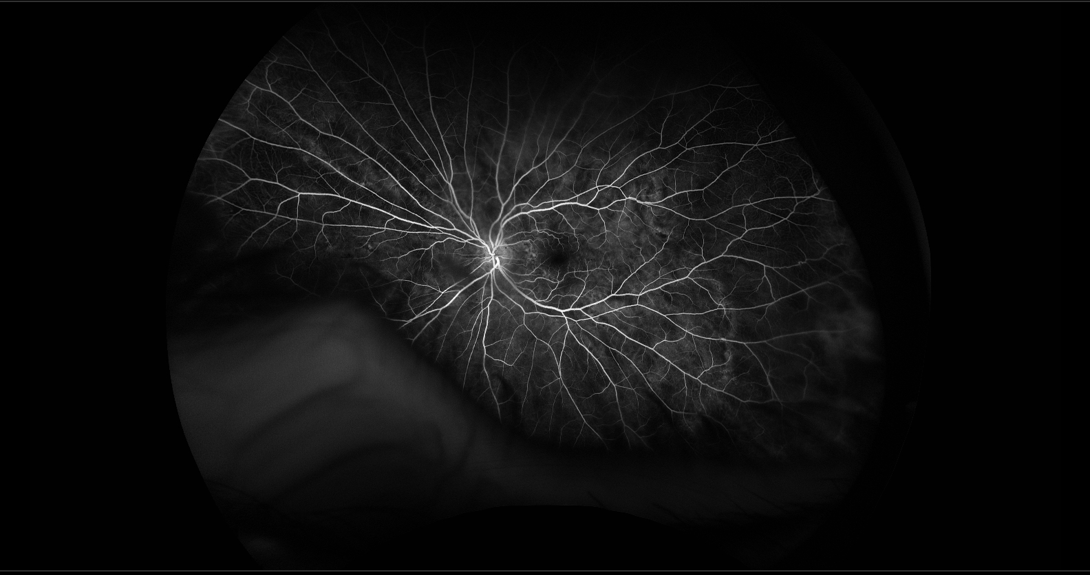
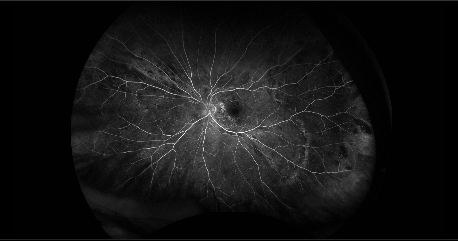
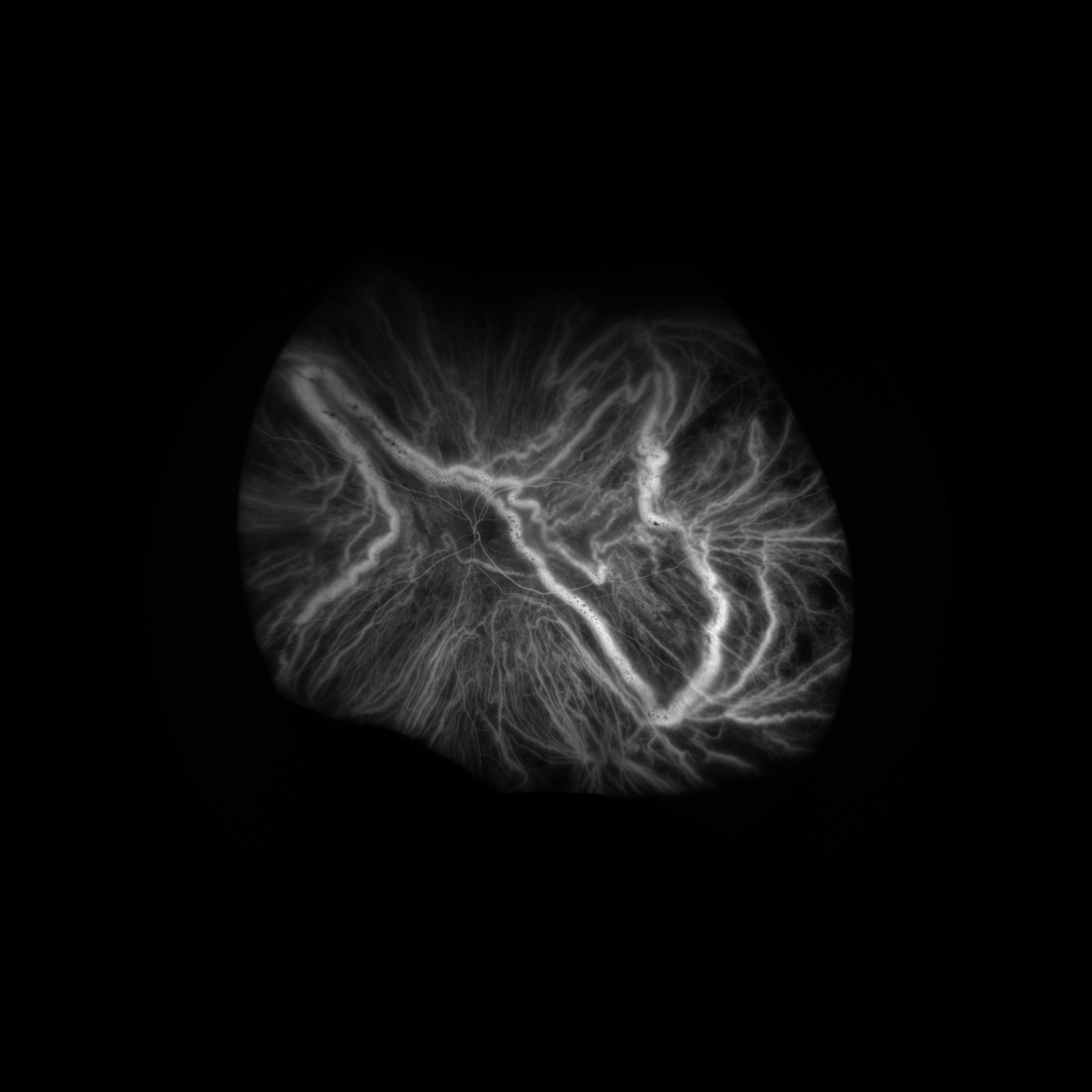
Imaging studies of the right eye are unremarkable. The fundus photograph of the left eye demonstrates a large, irregular, temporal linear loop of hypopigmentation with focal areas of hyperpigmentation. Fundus autofluorescence reveals linear and punctate hyperautofluorescence along portions of the loop, as well as two subtle linear hyperautofluorescent lesions nasal to the disc, indicating retinal pigment epithelium (RPE) stress.
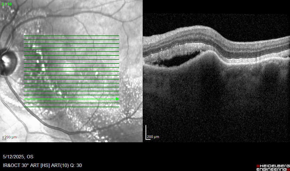
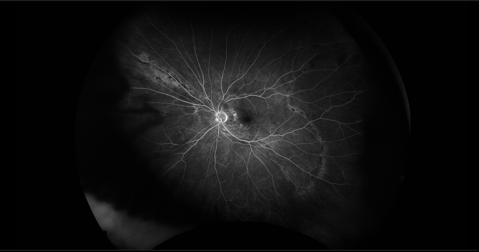
The red-free image (left of the OCT image) shows numerous hyperreflective dots both along and adjacent to the linear lesion, consistent with reflective exudative material. OCT demonstrates subretinal fluid with elongated photoreceptors, suggesting chronicity, as well as elevation of the retina and RPE by the choroidal macrovessel, disruption of the overlying choriocapillaris, and choroidal lucency corresponding to the vessel. Fluorescein angiography shows normal filling times and hyperfluorescence of the temporal loop and a linear lesion superonasal to the disc without late leakage. Indocyanine green angiography reveals hyperfluorescence corresponding to the irregular lesions that also exhibit hyperautofluorescence.
A choroidal macrovessel is a rare, abnormally dilated choroidal blood vessel. It is usually asymptomatic and is most often identified incidentally during routine fundus examination in middle or late adulthood. Although its etiology is unknown, it is generally considered a congenital developmental anomaly. In some cases, retinal elevation and subretinal fluid may produce metamorphopsia or decreased vision. Indocyanine green angiography is particularly helpful in establishing the diagnosis, as demonstrated in this case. No proven treatment exists for symptomatic patients, although anti-VEGF therapy has been attempted.
References
Gallo B, de Silva SR, Mahroo OA, et al. Choroidal macrovessels: multimodal imaging findings and review of the literature. Br J Ophthalmol. 2022;106(5):568-575.
Pichi F, Morara M, Veronese C, et al. Multimodal Imaging in Choroidal Macroaneurysms. Retina. 2013;33(7):1568-1577.
Mopuru R, Liu TYA, Arevalo JF. Choroidal Macrovessel Diagnosed on Multimodal Imaging, including Swept-Source Optical Coherence Tomography Angiography. Case Rep Ophthalmol. 2022;13(2):215-219.