Case Of The Month | July 2025

Case of the Month
July 21, 2025

The Case

The Case:

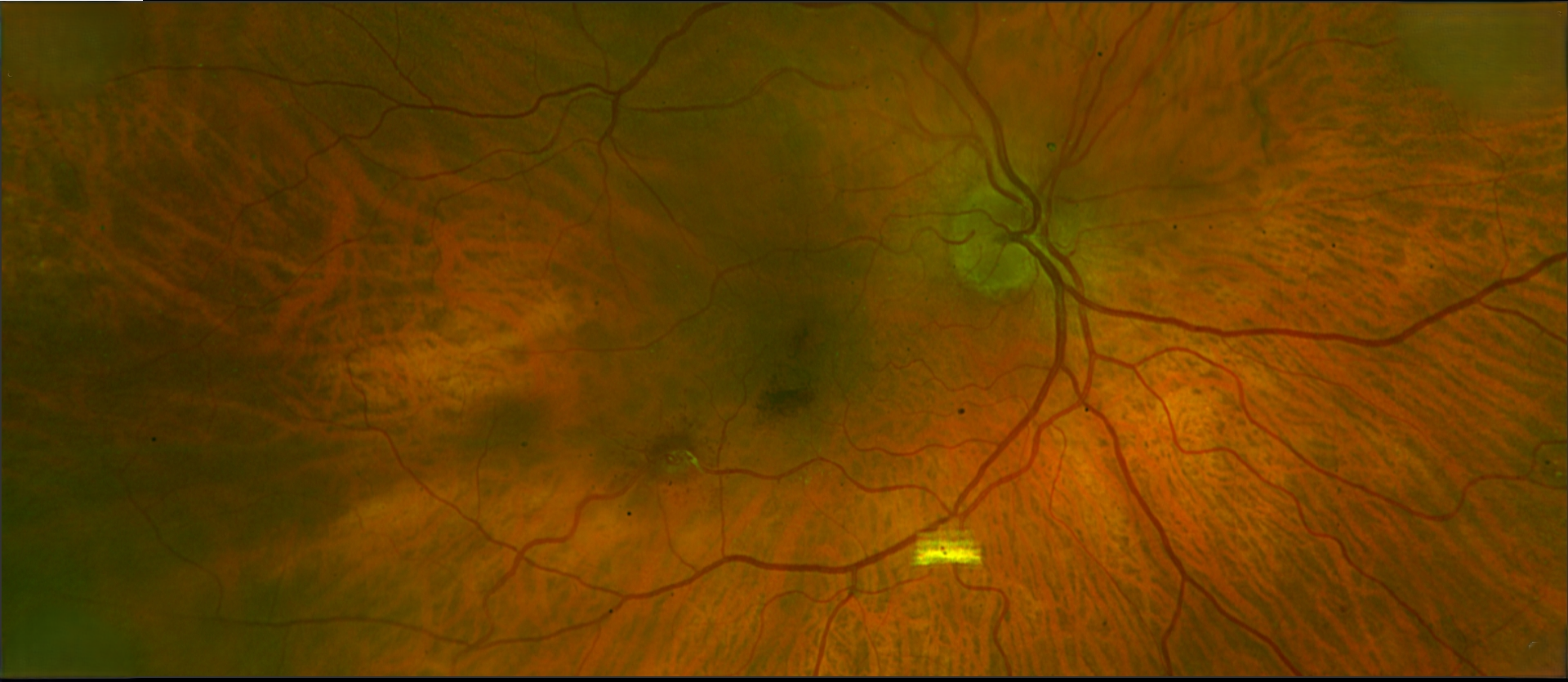
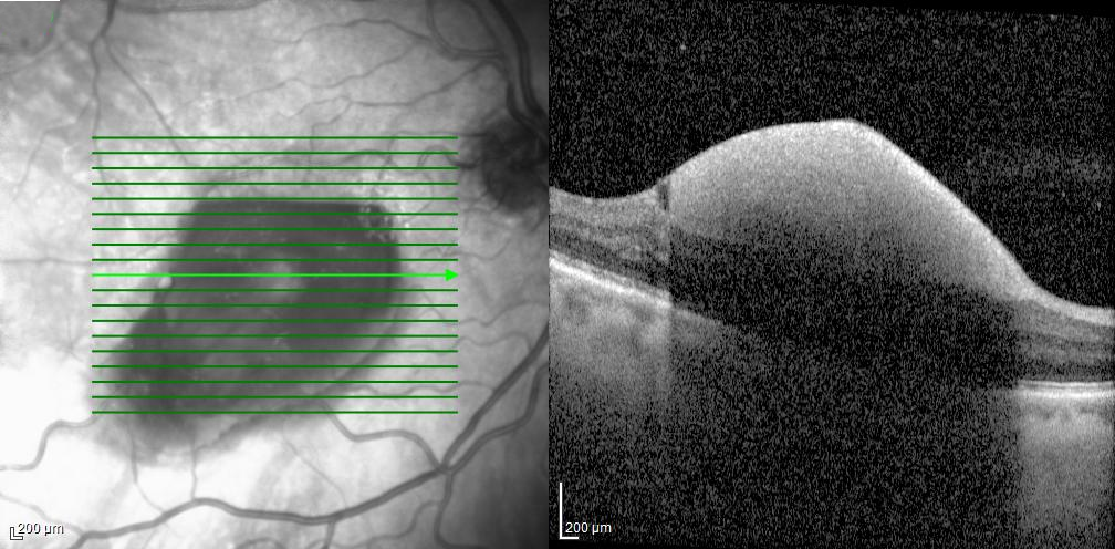
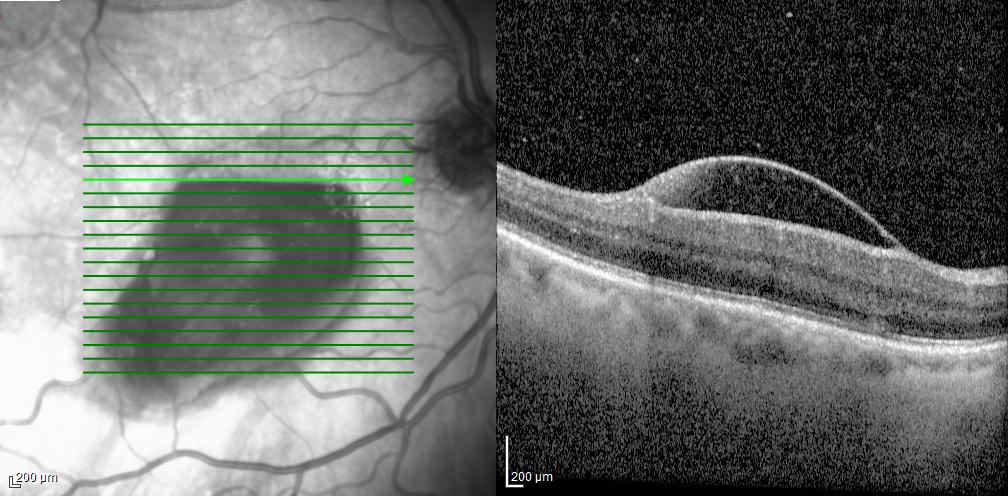
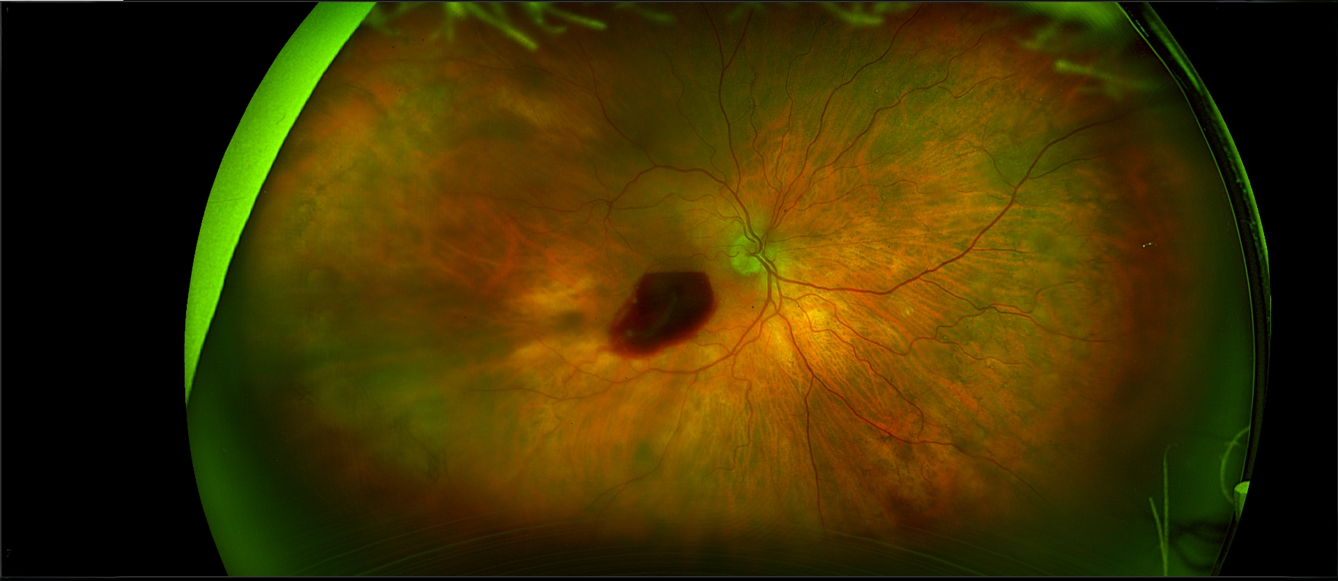
The patient was a 71-year-old woman with a sudden loss of vision in the right eye 4 days prior to our seeing her. She said that she recently had a cold and had been coughing heavily. The past medical history was unremarkable. The visual acuity was 20/200 OD and 20/40 OS. The anterior segment examination was remarkable for pseudophakia without significant capsular opacity in both eyes. The posterior segment examination was remarkable for a superficial hemorrhage in the right macula. There was no posterior vitreous detachment in the right eye. The left macula and the periphery of both eyes was unremarkable. What are the diagnostic considerations? What treatment, if any, would you recommend?

Photo OD 6/5

Answer:

The history and initial presentation suggested Valsalva retinopathy. Other diagnostic considerations include retinal artery macroaneurysm, a choroidal neovascular membrane (which can have blood breakthrough into the preretinal space), and trauma. The horizontally layered superior border indicated that the blood was located in the subhyaloid space. In contrast, blood under the internal limiting membrane (ILM) is generally diffuse and usually does not layer. Blood in the subhyaloid space tends to clear more readily than blood under the ILM. In our patient, there was substantial clearing of the blood over the following two months, and this revealed a retinal artery macroaneurysm inferotemporal to the fovea as the source of the blood.


Retinal artery macroaneurysms can lead to hemorrhaging in the preretinal, intraretinal, and subretinal spaces. Very large preretinal hemorrhages sometimes require surgical evacuation. Another approach has been to create a hole in the subhyaloid space and/or the ILM with a Nd:YAG laser, allowing the blood to leak into the vitreous where it is more readily absorbed.


Reference


IijimaH, Satoh S, Tsukahara S. Nd:YAG laser photodisruption for preretinal hemorrhagedue to retinal macroaneurysm. Retina 1998;18:430-4.

Case Photos

Click the Images below to enlarge
Photo OD 6/5

Answer:

The history and initial presentation suggested Valsalva retinopathy. Other diagnostic considerations include retinal artery macroaneurysm, a choroidal neovascular membrane (which can have blood breakthrough into the preretinal space), and trauma. The horizontally layered superior border indicated that the blood was located in the subhyaloid space. In contrast, blood under the internal limiting membrane (ILM) is generally diffuse and usually does not layer. Blood in the subhyaloid space tends to clear more readily than blood under the ILM. In our patient, there was substantial clearing of the blood over the following two months, and this revealed a retinal artery macroaneurysm inferotemporal to the fovea as the source of the blood.


Retinal artery macroaneurysms can lead to hemorrhaging in the preretinal, intraretinal, and subretinal spaces. Very large preretinal hemorrhages sometimes require surgical evacuation. Another approach has been to create a hole in the subhyaloid space and/or the ILM with a Nd:YAG laser, allowing the blood to leak into the vitreous where it is more readily absorbed.


Reference


IijimaH, Satoh S, Tsukahara S. Nd:YAG laser photodisruption for preretinal hemorrhagedue to retinal macroaneurysm. Retina 1998;18:430-4.

Genetics & AREDS Essay
Return to News & Events