Answer
When the patient was seen 1 ½ years later, the visual acuity was 20/20-1 J7 OD. The intraretinal hemorrhages and retinal whitening had resolved. The patient had not noticed any significant change in vision.
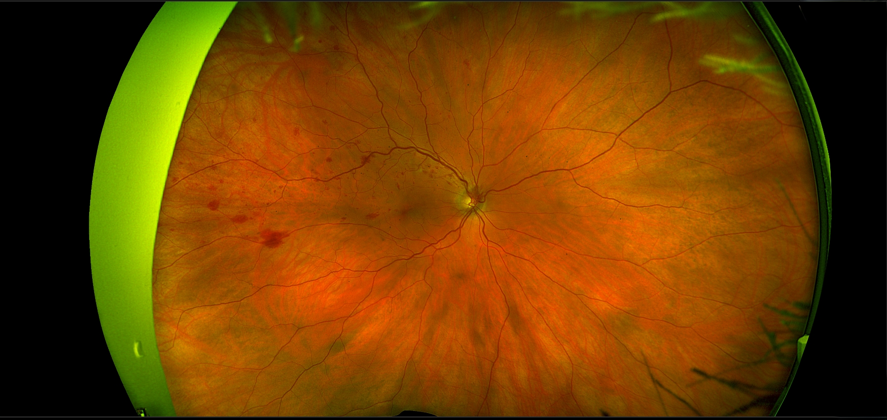
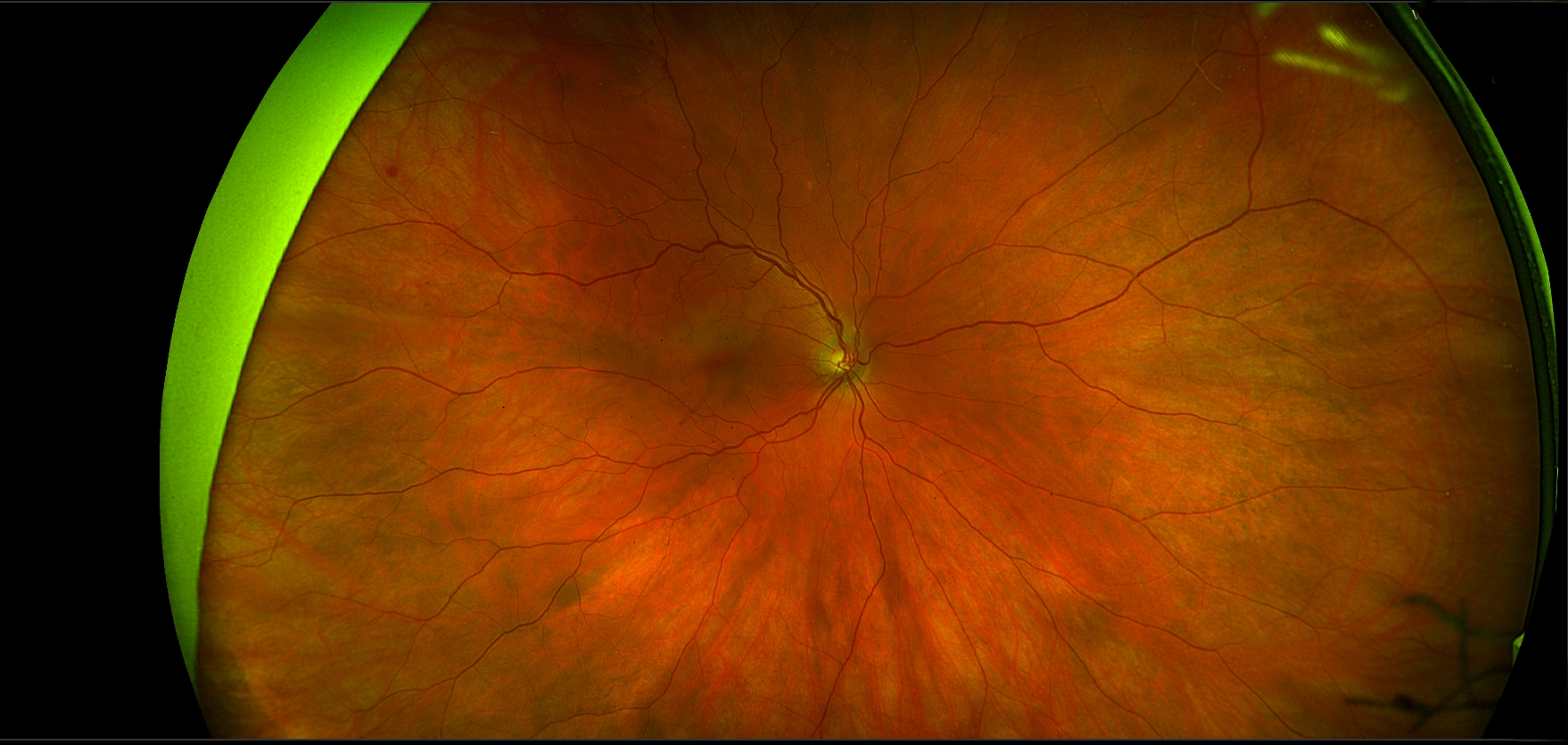
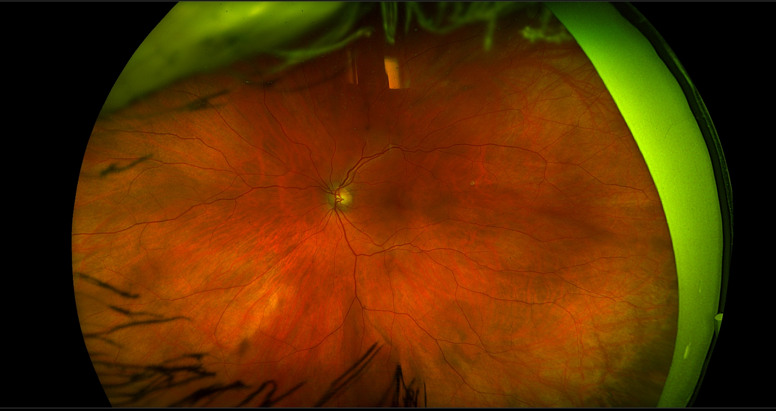
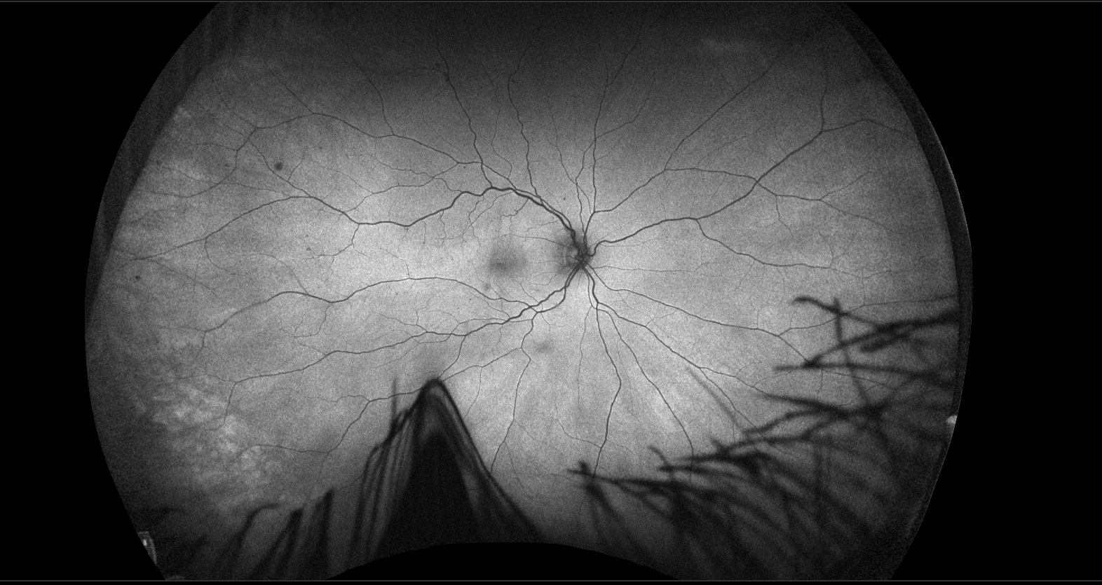
The initial fundus photographs showed several small hemorrhages and one larger hemorrhage in the right eye, as well as retinal swelling in the superior half of the macula. One month later, the hemorrhages had increased substantially, while the retinal whitening had decreased. The fundus photographs revealed dilation of the superotemporal retinal vein and greater venous tortuosity in the superotemporal quadrant than in the inferotemporal quadrant at both the initial presentation and one month later. The degree of venous tortuosity did not change significantly after one month, despite the substantial increase in hemorrhages. The autofluorescence image was remarkable only for the presence of hemorrhages.
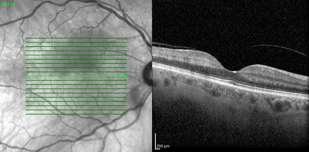
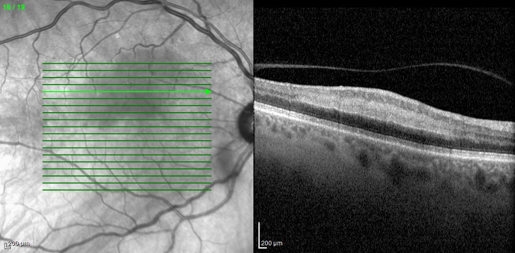
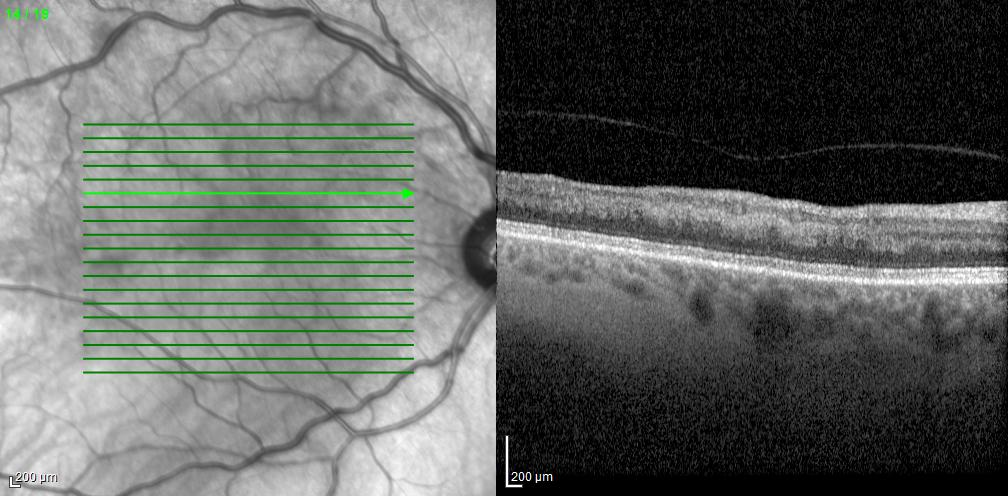
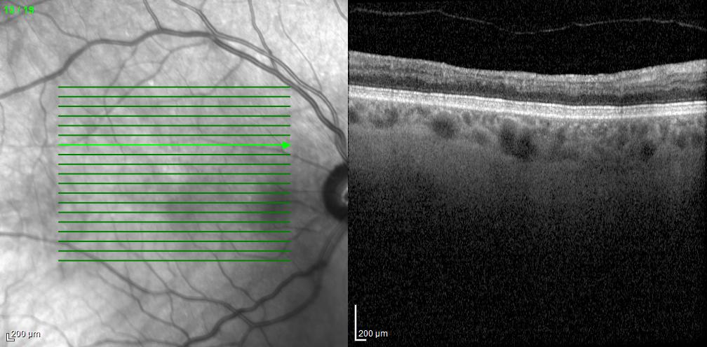
OCT revealed increased reflectivity in the middle retinal layers, consistent with edema. One month later, the reflectivity had decreased significantly. The intraretinal edema gradually resolved, and 7 months later there was no residual increased reflectivity or edema. However, there was retinal thinning and disruption of the middle retinal laminations in the superotemporal macula.
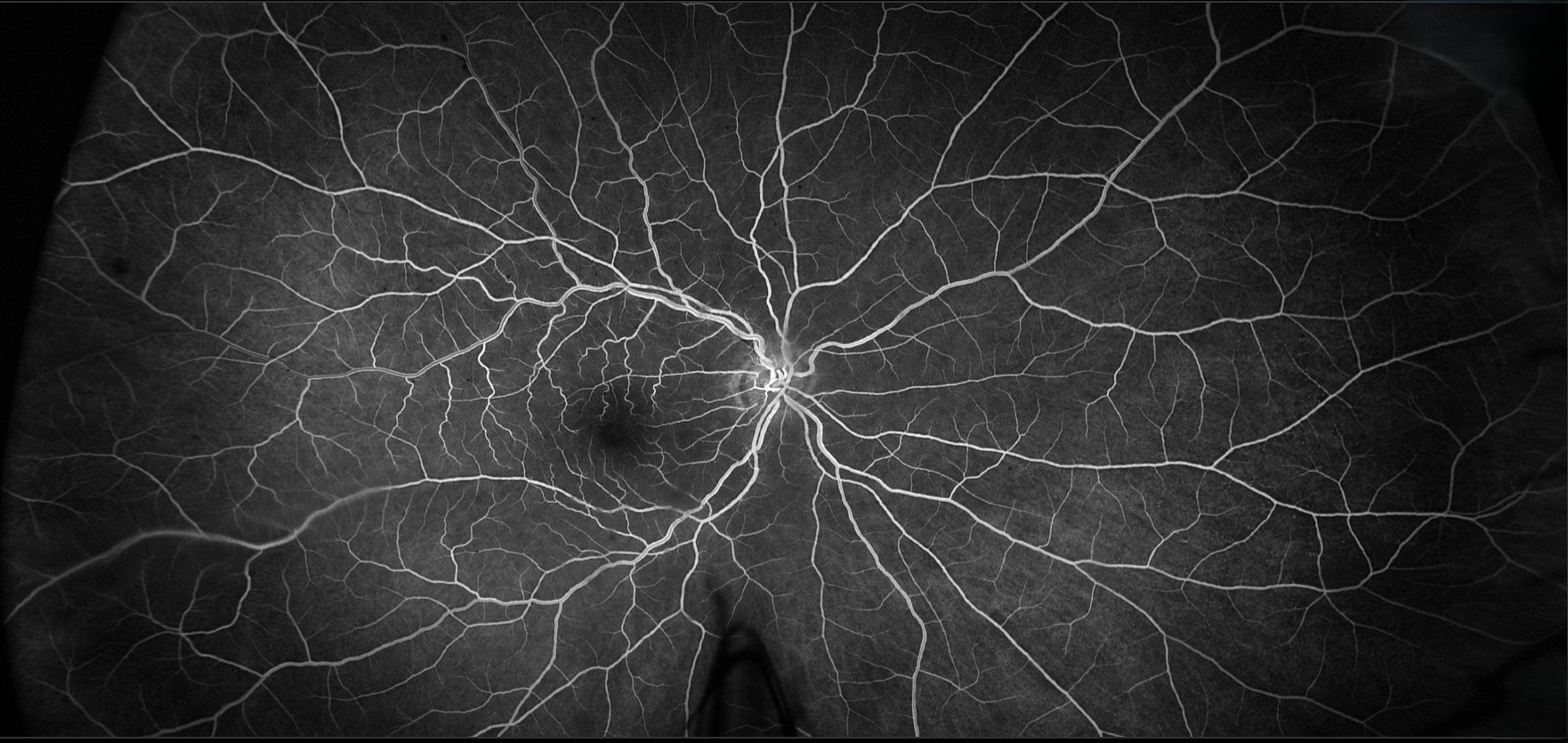
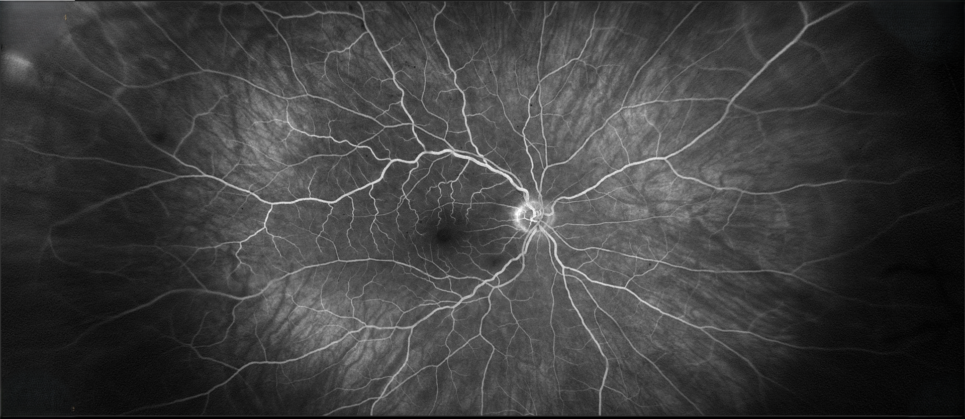
Fluorescein angiography at presentation revealed slightly delayed superotemporal venous filling and moderate tortuosity of the superotemporal veins compared with the inferotemporal veins. There was no significant leakage.
This patient had a branch retinal vein occlusion (BRVO) with an unusual clinical course. BRVOs can induce paracentral acute middle maculopathy (PAMM), and cases of BRVO associated with PAMM without significant intraretinal hemorrhaging have been reported. PAMM results from impaired blood flow in the intermediate and/or deep retinal capillary plexuses, producing edema that tends to be most prominent in the inner nuclear layer. There are many possible causes, including venous occlusive disease, arterial occlusive disease, diabetic retinopathy, and hypertensive retinopathy.
Because there was no cystoid macular edema, we elected to observe the patient closely. She did not notice any change in vision over time, which is consistent with the typical clinical course. Patients with PAMM often experience decreased vision and a permanent scotoma, although the visual symptoms may improve slightly after the initial presentation.
References
Fumi D, Ruggeri F, Fasciolo D, Antonello, E, Burtini G, Abdolrahimzadeh S. Paracentral acute middle maculopathy (PAMM) in ocular vascular disease – what we know and future prospects. Vision 2025; 9:19.
Casalino G, Williams M, McAvoy C, Bandello F, Chakravarthy U. Optical coherence tomography angiography in paracentral acute middle maculopathy secondary to central retinal vein occlusion. Eye 2016, 30, 888–893.