Answer
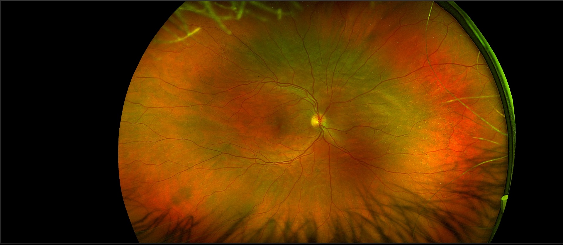
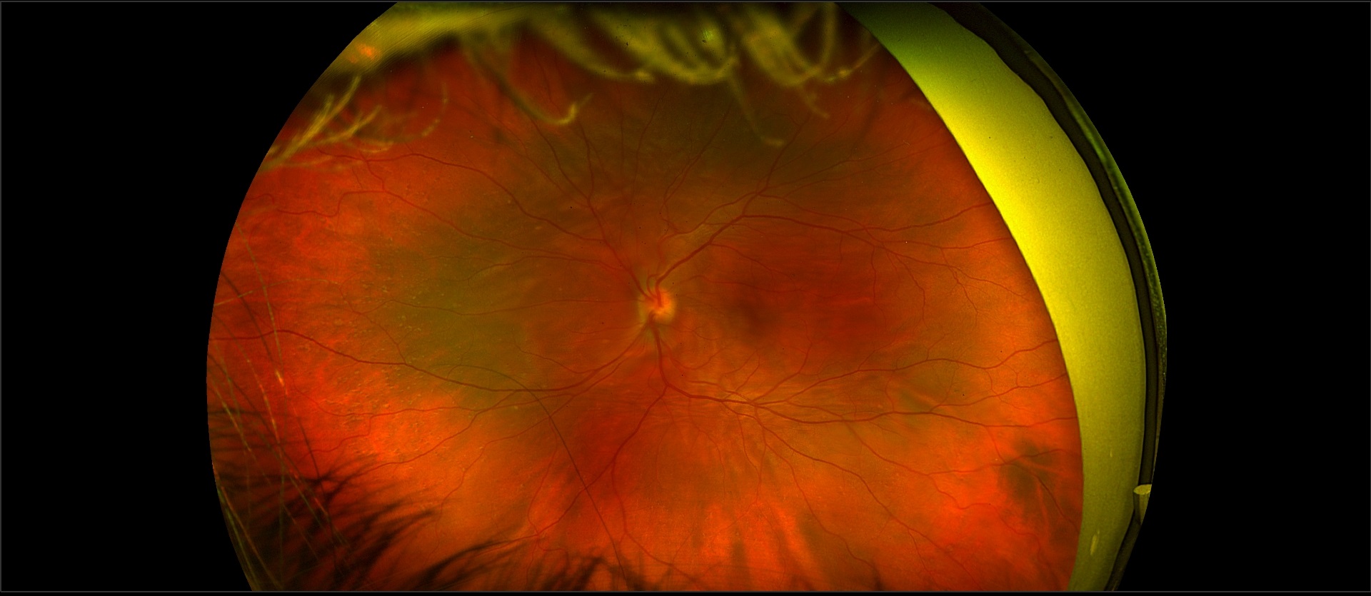
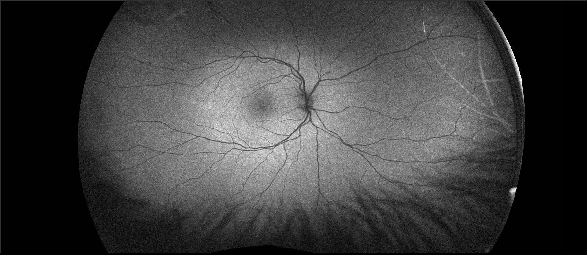
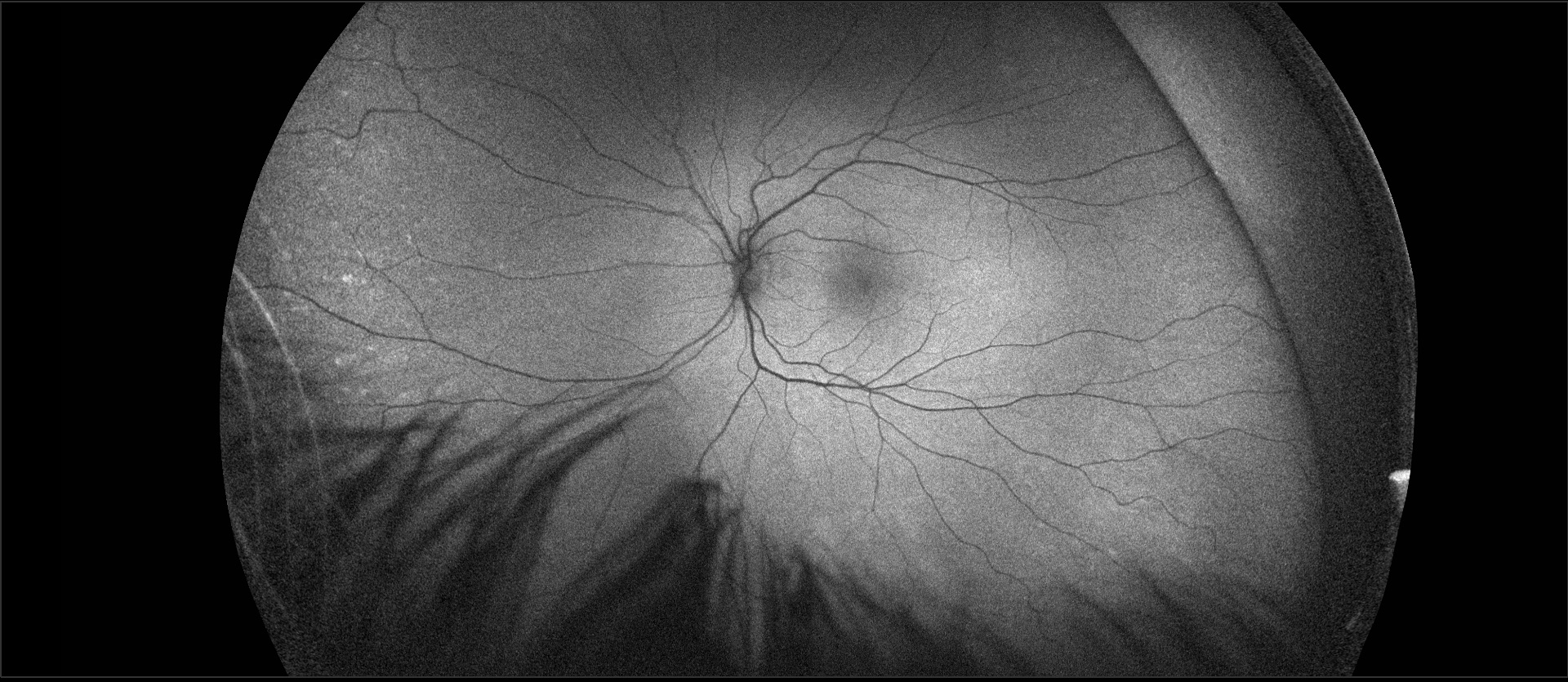
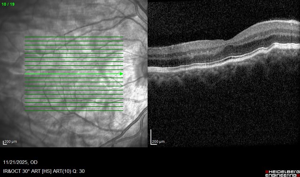
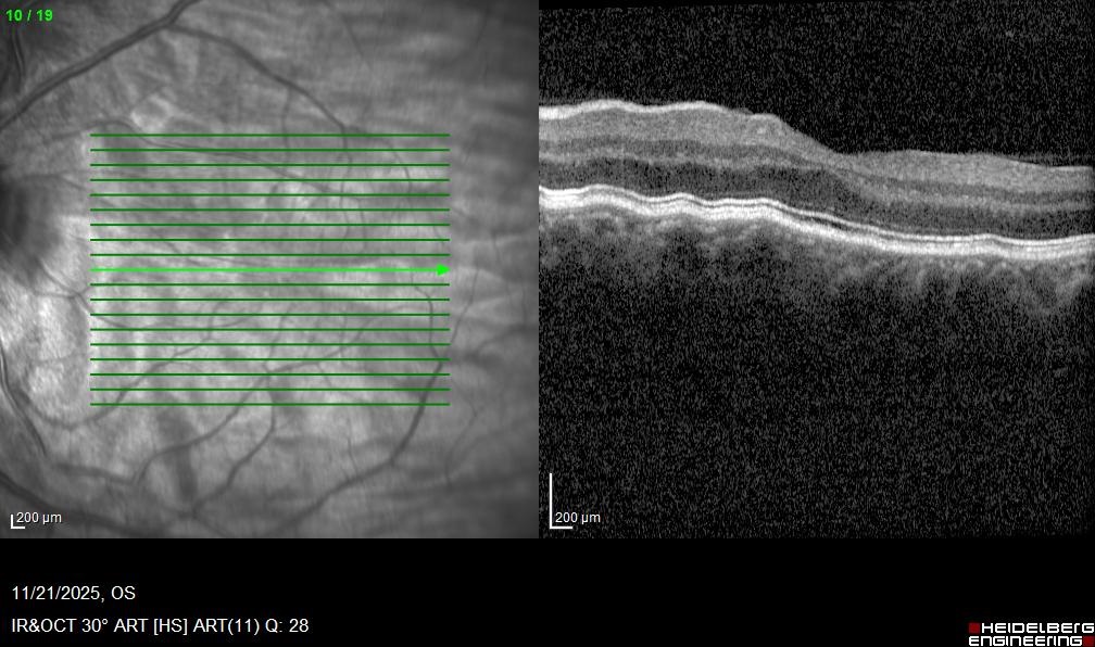
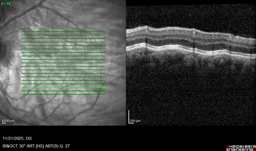
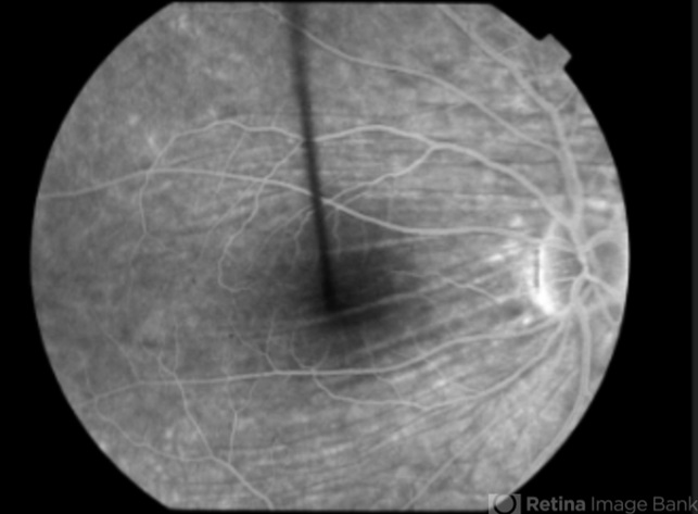
The transient blurred vision was likely related to contact lens use. Fundus examination and imaging studies revealed bilateral choroidal folds. The folds are visible in the color photographs and in the red-free images accompanying the OCTs, but they are not well visualized on autofluorescence imaging. The OCTs demonstrate the characteristic undulations of the choroid, with normal choroidal thickness and unremarkable overlying retinal structures.
In this condition, fluorescein angiography typically shows hyperfluorescence at the peaks of the folds and hypofluorescence in the valleys. In contrast, it does not demonstrate retinal folds. Fluorescein leakage may be seen in processes underlying retinal folds, such as an epiretinal membrane.
Bilateral choroidal folds are most commonly idiopathic. In this patient, they are associated with high hyperopia.
The differential diagnosis for unilateral choroidal folds is much broader and includes orbital tumors; orbital inflammatory conditions such as pseudotumor; orbital infections; thyroid orbitopathy; choroidal tumors; choroidal neovascular membranes; inflammatory conditions such as Vogt–Koyanagi–Harada syndrome; pachychoroid spectrum disorders (particularly peripapillary pachychoroid syndrome); uveal effusion syndrome; retinal vascular occlusions; retinal detachment; posterior staphyloma; and hypotony.
Cheng JY, Hsu D, Feo A, et al. Choroidal folds: A review and update of new and old etiologies. Survey of Ophthalmology 2026; 71(2):405-422.